English


Endoscopic Discectomy to Treat Herniated Disc
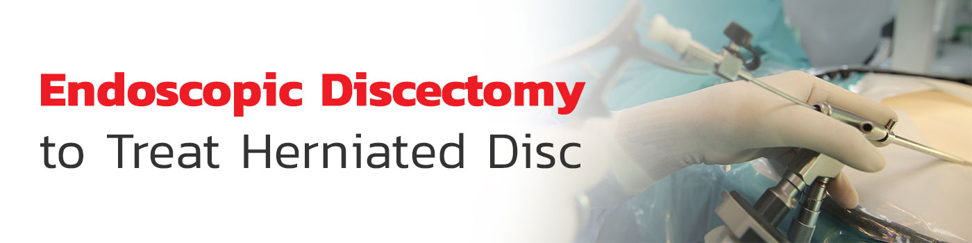
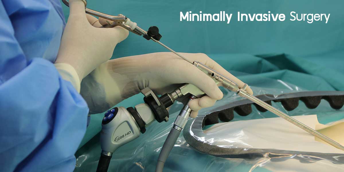
Endoscopic discectomy
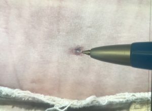
An endoscope is a small surgical instrument which is shaped like a pipe with a 5 millimeter diameter. At one end is lens while the inside is a fiber optic to show the image of the affected area. There is space to insert equipment such as a laser or other special treatment equipment to help treat areas not visible to the naked eye.
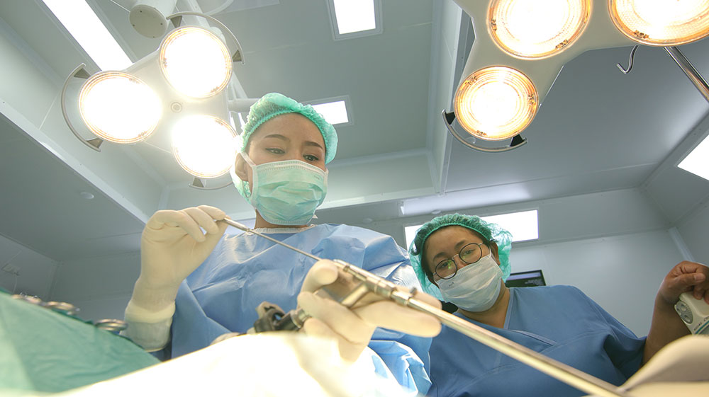

After the patient is administered anesthetic, the physician will make a 0.5 centimeter incision on the flank or the back and insert the endoscope through the incision to the affected area to treat the depressed nerve. The physician will not have to cut out any back muscle to reach the affected area.
The endoscope will help the physician see the nerve more clearly as it can penetrate more deeply and magnify the view. This will make it easy for the physician to remove the specific part of the spinal disc, the spinal joint, or the tendon which is depressing the nerve. The procedure takes only 15-30 minutes and the patient will be able to get up and walk post-surgery.
Benefits
- Only a 0.5 centimeter incision so less pain from the incision wound post-surgery
- Low risk of infection
- Lessens the damage on healthy tissue surrounding the incision wound
- Speedy recovery post-surgery and discharge within 24 hours
- Blood transfusion not necessary due to limited loss of blood
- Lower cost than normal surgery because the special equipment means less injury and less recuperation needed at the hospital
Disadvantages
- Only applicable for cases where the spinal disc hasn’t dislocated much; cases where the spinal disc has dislocated far requires the insertion of screws to fix the spinal disc in place
- Must be conducted by an experienced physician for a successful result and limited complications