English


Do you need to undergo X-ray or MRI test for your back pain?
Many people would have wondered what is the difference between X-ray and MRI since both are used for diagnosing disorders in our bodies. Well, today we have the answer for you.

X-RAY
X-ray is an image of the body many people would be familiar with. This image is made by electromagnetic radiation that passes through the damaged area and a detector on the other side picks up the beams and turns it into a shadowgraph. The image is shown in different shades of black and white depending on how much the X-ray can pass through the object. The X-ray appears white if the beams pass through dense parts of the body (like bones), while other parts (like soft tissues) that allow more beams to pass through appears dark grey.
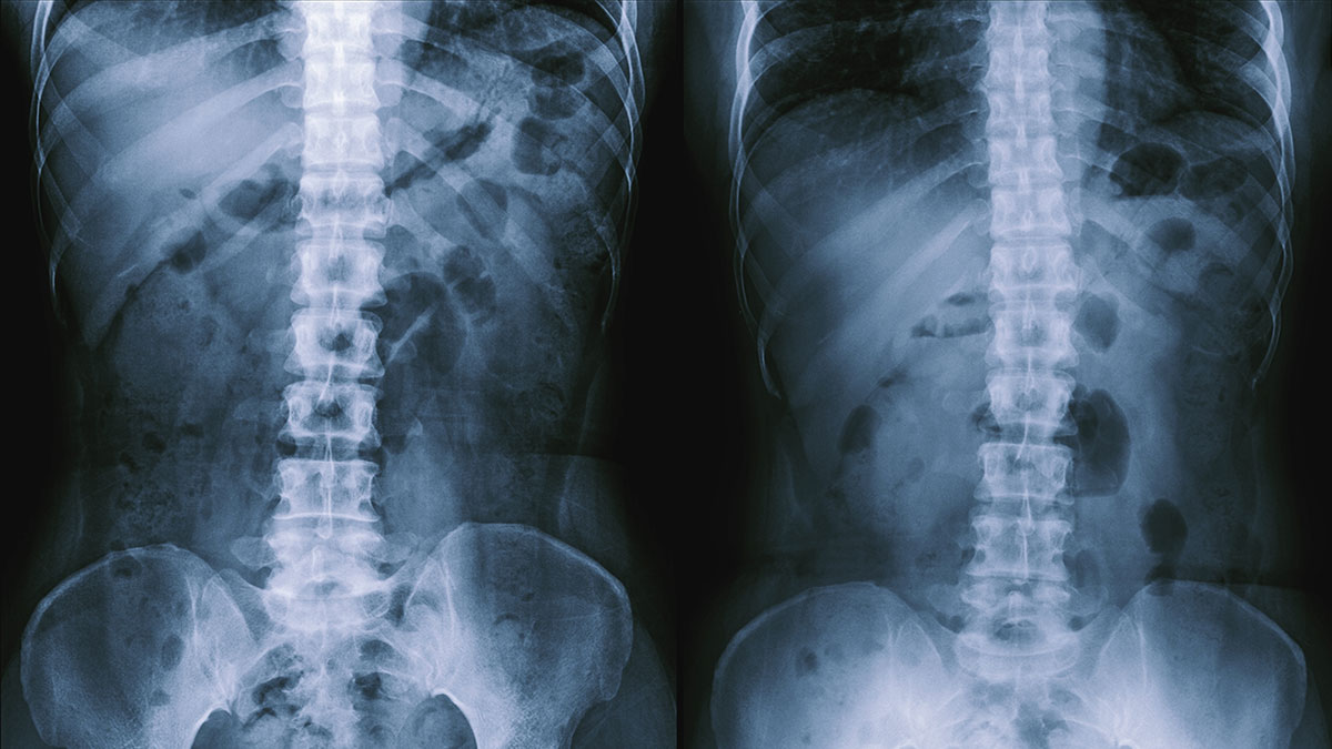
Here is an X-ray of the spine, you can see the vertebrae as well as the transparent parts between each vertebra which are intervertebral discs.
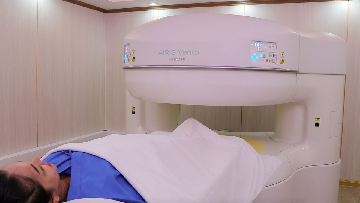
MRI
Magnetic Resonance Imaging, MRI, uses magnetic fields and radio waves to create a clear detailed image with the ability to highlight problems in soft tissues, regardless of the density of tissues. Hence, it can detect various issues of the brain, ligaments, tendons, nerve, muscle, spinal cord, and skeleton. To put it simply, MRI allows the doctor to see through all the parts of the body.
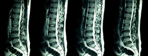
This is a picture of the spine, whereby the MRI allows clearer vision of elements of the spine such as inter-vertebral disc.
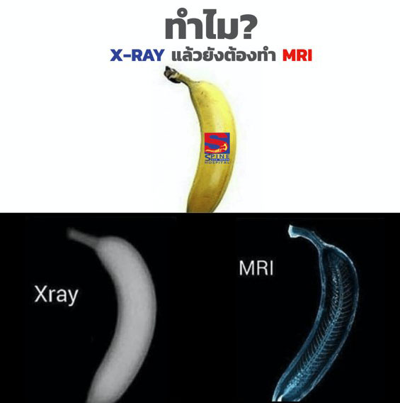
Why are both X-ray and MRI recommended?
In case you have spine issues, feel the back pain radiating to the legs, or unable to find the cause of the pain, you need both X-ray and MRI to allow the doctor to make an accurate diagnosis. X-ray produces the clear image of spinal structure, while MRI allows the doctor to see disorders of the discs.
It is important to have a specialist doctor or radiologist to read and interpret the scan to avoid inaccurate diagnosis. At S spine and Nerve Hospital, we have specialty doctors who can make an accurate diagnosis from X-ray and MRI. Plus, we are the specialist hospital in spine and nervous system.